Ophthalmic toxicology
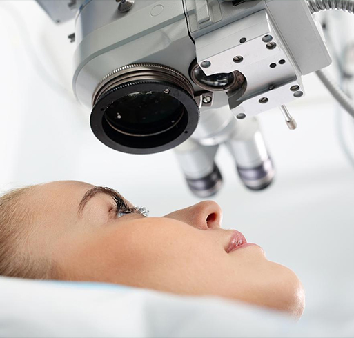
To evaluate the safety and effectiveness of ophthalmic drugs, and to evaluate the effects of drugs on the eye after systemic administration. He is good at various intraocular drug administration in different species of animals, and has established experimental animal models of corneal neovascularization, corneal epithelial injury and cataract. With advanced eye examination instruments, such as slit-lamp, tachymeter, fundus camera, OCT, electroretinogram, and ophthalmic operating microscope, it can perform complete live animal eye examination and provide support for preclinical research of ophthalmic drugs.
Category: Non-clinical safety evaluation
Medicines for human purposes
I. Introduction
To evaluate the safety and effectiveness of ophthalmic drugs, and to evaluate the effects of drugs on the eye after systemic administration. He is good at various intraocular drug administration in different species of animals, and has established experimental animal models of corneal neovascularization, corneal epithelial injury and cataract. With advanced eye examination instruments, such as slit-lamp, tachymeter, fundus camera, OCT, electroretinogram, and ophthalmic operating microscope, it can perform complete live animal eye examination and provide support for preclinical research of ophthalmic drugs.
Ii. Main research content
1. Non-clinical studies of ophthalmic drugs:
Safety evaluation, pharmacodynamic evaluation, pharmacokinetics/toxokinetics (eye circulation)
2. Animal species:
Rodents (large, mouse), rabbits, dogs, non-human primates
3. Method of administration:
Topical ophthalmic use: intraconjunctival capsule administration
Intraocular injection: anterior chamber injection, vitreous cavity injection, subconjunctival injection, retrobulbar injection
Systemic administration: intravenous, oral, intramuscular, subcutaneous
4. Eye disease model:
Clinical disease
Molding method
Animal germline
Corneal neovascularization
Corneal suture induction
New Zealand white rabbit
Alkali burn induction
Corneal epithelial injury
Corneal epithelial cell curettage induction
New Zealand white rabbit
cataract
Induction by subcutaneous injection of sodium selenite
SD rat
Retinal melanosis
Retigabine induction
Long Evans rat
5. Live eye examination:
Slit-lamp examination and corneal fluorescein sodium staining examination
Direct/indirect ophthalmoscopy
Fundus photography FP
Fundus fluorescence imaging FFA
Optical coherence tomography OCT
Infrared reflection imaging IR
Fundus autofluorescence scanning BAF
Intraocular pressure IOP
Electroretinogram ERG
Visual evoked potential (VEP)
